Where measurement happens
The region of interest is not a neutral container around a feature calculation.
It states which tissue the measurement is meant to represent. Intratumoral,
peritumoral, multi-region, and dose-defined measurements can use the same image
and extractor while answering different questions. The region is therefore part
of the operational definition:
feature = f(image, region definition, geometry, preprocessing, implementation).
The region is a hypothesis
An intratumoral measurement tests whether the internal architecture of the lesion contains the relevant signal. A peritumoral measurement tests a different idea: that the interface with surrounding tissue carries information. A multi-region measurement proposes that compartments provide complementary information. In a radiotherapy setting, a dose-stratified region can encode a hypothesis about where dose meets tissue and how that relates to toxicity. None of these regions is the default biological truth. Each is a design choice whose construction, exclusions, and intended interpretation should be stated before results are compared.
This framing also clarifies why a mask can be technically valid and scientifically wrong for a question. A perfectly aligned ring around a tumour still measures the tissue included by the ring, not an abstract quantity called “the tumour”. If the region changes, the estimand changes with it.
Rings live in physical space
A peritumoral ring can be constructed by dilating a tumour mask and subtracting the original mask; erosion can define a core. The width must be expressed in millimetres of patient space or as a physical-distance criterion on a declared grid. “A three-voxel ring” is not portable across scans: three voxels at 1 mm spacing span a different physical distance from three voxels at 5 mm spacing. Dilation on raw indices across heterogeneous geometries therefore creates different regions while giving them the same name.
Where the lesion sits also changes what the ring contains. A ring around a peripheral lung nodule may include lung parenchyma, vessels, or chest wall; a ring around a central lesion may include bronchi or mediastinal fat. If the hypothesis concerns one tissue, exclusion rules may be appropriate, but they are part of the definition and must be reported. They are not a universal repair that can be added after seeing which region performs best.
A published induction-therapy example illustrates the specificity of such a rule: its methods describe a 3 mm three-dimensional dilation minus the tumour, with areas next to the chest wall and vertebral bodies excluded by radiologists. That is a clear operational definition for that study. It does not establish that 3 mm is a preferred width or that the same exclusions are appropriate for every anatomy or endpoint.
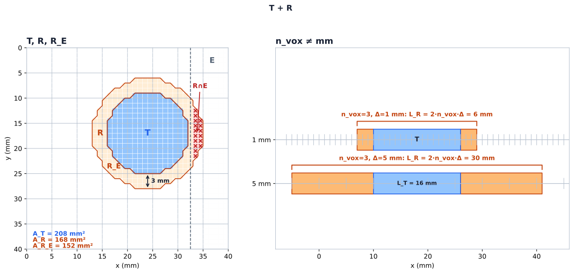
Synthetic geometry, not patient data: the areas and lengths characterise this construction only. The figure does not show that a ring measures tumour biology, that 3 mm is a preferred width, or that clipping is optional — the exclusion rule is part of the measurement definition.
Region choices change sensitivity and uncertainty
Changing a region changes more than its label. A boundary perturbation moves the inner edge of a ring and can also move its outer edge. A fixed physical width becomes a different voxel count if the target grid changes. The same ring can be a large addition around a small lesion and a modest addition around a large one, so region sensitivity interacts with lesion size and with partial-volume composition. These are reasons to characterize region-related variation, not reasons to assume that one region is always superior.
The biological interpretation is conditional as well. A peritumoral feature may capture surrounding anatomy or acquisition context rather than the intended tumour process. A background-region control or a comparison with tumour-only features can probe whether the added region contributes information beyond size and local context. These controls probe incremental information and alternative explanations; they do not identify a mechanism by themselves.
Width selection is a multiplicity problem
Trying several widths, several region combinations, and several feature sets is a selection process. Reporting the best width as though it had been fixed in advance turns a researcher degree of freedom into an apparent biological constant. The fact that published studies in different cohorts and endpoints have reported different preferred widths is therefore unsurprising; it is not evidence that one width travels across settings.
A region should be prespecified when the scientific rationale supports one. If a study explores widths, the exploration should be declared as part of the analysis, compared with a prespecified baseline relevant to the estimand, and handled as a source of selection uncertainty rather than hidden in the final label. The same principle applies to habitat regions defined by clustering: the clustering rule is part of the region definition and must be frozen and reported.
Once the region has been defined, the next question is how two measurements of it agree. The following section separates linear association from agreement with the line of identity.
Sources
- Induction-therapy response radiomics with 3 mm dilation minus tumour. BMC Cancer. 2025. doi:10.1186/s12885-025-13804-x.
© 2026 Adrian Tkacz · BioMedical-IT
Original text and figures are licensed under the Creative Commons Attribution-NonCommercial 4.0 International License (CC BY-NC 4.0), unless otherwise noted. Please credit Adrian Tkacz / BioMedical-IT and link to the original source. Third-party materials retain their respective rights and licenses.