Reconstruction as a Task-Dependent Transformation
The reconstructed image is not the biological object
A CT reconstruction is an operation that turns acquired measurement data into an image representation. It can change the visible noise, resolution, texture, and local contrast without changing the patient or the underlying anatomy. The mistake is to treat a difference between images as a difference in biology. The more precise statement is that the same biological object can produce different image evidence, and a measurement or model operating on that evidence can therefore change.
Filtered back-projection, iterative reconstruction, and learned reconstruction are families of reconstruction approaches, not a universal progression from bad to good. Their behaviour depends on the acquisition, the reconstruction objective, the implementation, and the parameters used. An iterative method may trade one kind of noise pattern for another. A learned method may reduce a particular noise pattern while also changing edges or texture. Those are possibilities to test for a specified system, not signatures that can be assigned to every implementation in the family. Suppression and hallucination are failure possibilities to test in that specified system, not inherent signatures of learned reconstruction.
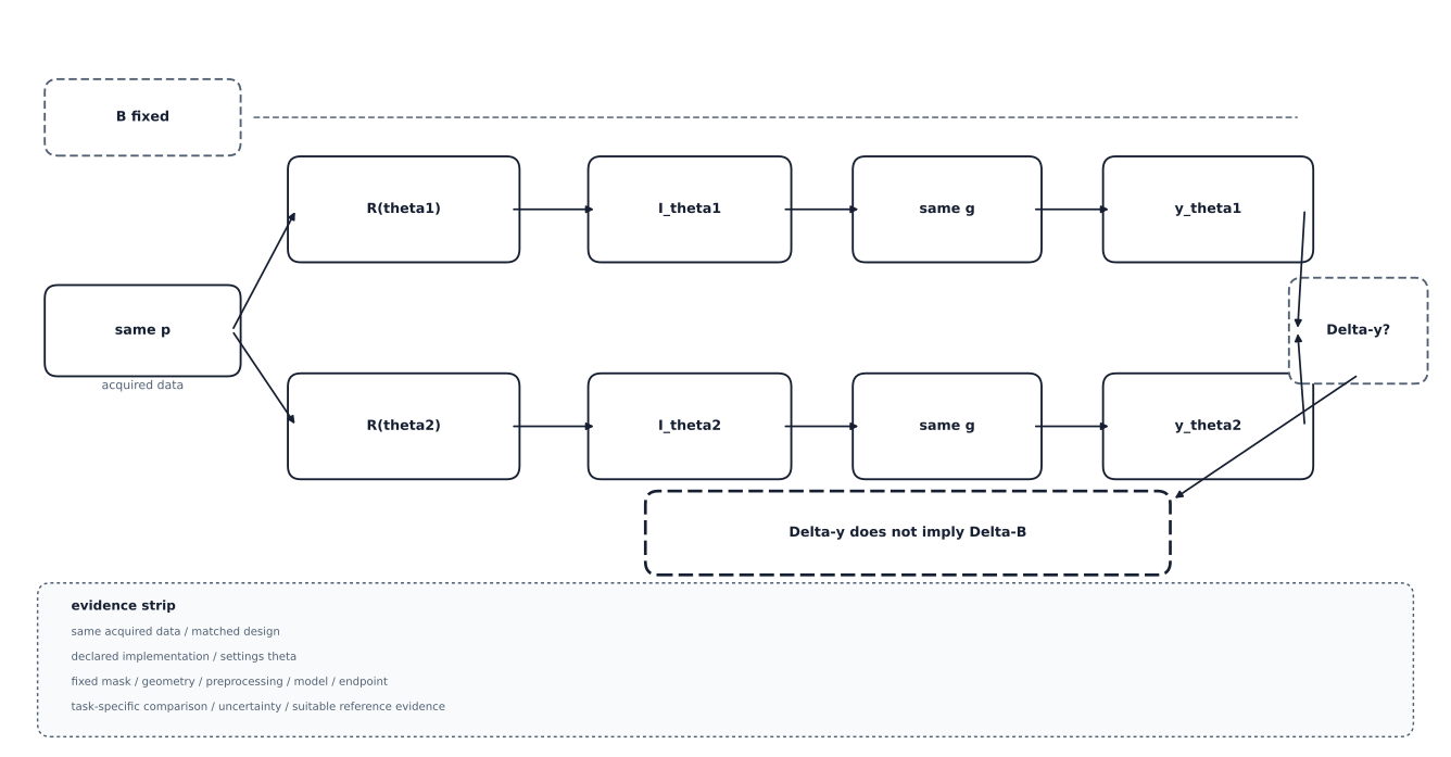
A controlled measurement-path schematic, not empirical evidence about FBP, iterative, or learned reconstruction. The same acquired data and unchanged biological object pass through two generic settings; only the declared reconstruction setting varies and the downstream function g is fixed. A difference between the resulting task outputs is evidence of reconstruction-dependent behaviour in that declared design, not evidence that biology changed and not a universal ranking of reconstruction families. Suppression or invention remains a failure possibility to test for a specified system, not an outcome asserted by this diagram.
Reconstruction is part of a measurement definition
For quantitative imaging, a useful mental model is y = f(image, mask, geometry, preprocessing).
The reconstruction is inside image: it helps determine the values from which
the measurement is calculated. If the mask, geometry, and feature implementation
are held fixed while the reconstruction changes, a changed feature value is
evidence of reconstruction-dependent measurement behaviour. It is not evidence
that the biological quantity itself changed.
This distinction matters for both cohorts and models. Two cohorts reconstructed under different conditions should not automatically be pooled as if their image values were observations of one identical measurement; whether pooling is defensible depends on the endpoint, the declared estimand, and sensitivity evidence. A model trained on one reconstruction setting and evaluated on another can face an input-distribution change. Whether that change matters, and whether adaptation is appropriate, depends on the task and the deployment claim. It cannot be settled by naming the reconstruction family alone.
The same reasoning applies to a learned reconstructor that produces a visually convincing image. If it removes a pattern that carried task information or adds a plausible pattern not supported by the acquired evidence, a downstream measurement can move without a corresponding biological change. This is a measurement and evidence question, not a claim about a universal failure mode of learned reconstruction.
Record the reconstruction and test the intended shift
A reconstruction-aware protocol records the reconstruction family and the relevant implementation details available for the study, such as vendor or strength settings, model or release information, kernel, slice representation, and the acquisition conditions that produced the data. The exact fields depend on the modality and object. If the reconstruction is not recorded, the available data may not identify how much of an observed difference belongs to reconstruction rather than site or biology; this is non-identifiability within the available data, not proof that the reconstruction caused the difference.
Pooling across reconstruction settings is not automatically forbidden. It is a design choice that needs a declared estimand and sensitivity evidence: a study may deliberately measure sensitivity to a reconstruction change, harmonise a declared difference, or restrict a model to one deployment regime. What is not defensible is to leave the change implicit and then attribute a resulting feature or performance difference to biology without examining the image-to-measurement path.
The deployment test should represent the reconstruction the model will meet. A biomarker or classifier developed on one setting is not automatically licensed for a different setting merely because both images depict the same anatomy. Frozen evaluation, task-specific measurement checks, and any recalibration or adaptation should be reported as separate parts of the procedure. The next section examines a more explicit transformation: producing an image for a target contrast or protocol that was not directly acquired.
© 2026 Adrian Tkacz · BioMedical-IT
Original text and figures are licensed under the Creative Commons Attribution-NonCommercial 4.0 International License (CC BY-NC 4.0), unless otherwise noted. Please credit Adrian Tkacz / BioMedical-IT and link to the original source. Third-party materials retain their respective rights and licenses.