Inversion recovery and signal suppression
A timing choice can create a null
Fat is often bright in common MR contrast, and that brightness can obscure a small signal difference in marrow or soft tissue. Suppressing fat is not a display trick applied after reconstruction. It is a sequence design choice that changes the longitudinal magnetisation before the signal is sampled. STIR is a particularly clear example because it uses a T1-dependent zero crossing.
An inversion-recovery (IR) sequence begins with a 180° inversion pulse that reverses the longitudinal magnetisation. The inverted magnetisation then recovers toward equilibrium. In an idealised model, it passes through zero during that recovery. If the sequence applies its excitation when the chosen tissue is at or near this zero crossing, little longitudinal magnetisation is available to create the subsequent transverse signal, and that tissue is nulled.
The interval from the inversion pulse to the excitation is the inversion time (TI).
In the idealised case of full equilibrium before a perfect inversion, the longitudinal
component can be written as M_z/M_0 = 1 − 2e^(−TI/T1), with a zero crossing at
TI = T1 ln 2. This equation is a teaching model, not a universal clinical setting:
finite TR, imperfect pulses, the readout, magnitude reconstruction, and the tissue’s
actual T1 all change the null point and the resulting contrast.
180° inversion pulse | | longitudinal magnetisation recovers through zero vChoose TI at the target tissue's zero crossing | vTarget tissue is suppressed while other tissues remain visibleSTIR and FLAIR use the same mechanism
STIR, or Short TI Inversion Recovery, chooses a short TI aimed at the T1 range of fat. This reduces fat signal while tissues whose longitudinal magnetisation is not near the same zero crossing retain more signal. Because the null is T1-based rather than chemically specific, STIR can also suppress other short-T1 tissues, including gadolinium-enhancing tissue, and should not be used after gadolinium when positive enhancement is the intended finding. In musculoskeletal and spinal imaging, this contrast is often used to make marrow or soft-tissue fluid-related abnormalities easier to see. The statement is about a contrast design, not a promise that every abnormality is bright or that every implementation produces the same appearance.
The same mental model explains FLAIR, Fluid-Attenuated Inversion Recovery. It uses a longer TI aimed at the T1 range of fluid such as CSF, reducing the normally bright fluid signal so other tissue differences can be inspected. STIR and FLAIR do not differ in the logic of nulling; they choose different target tissues and therefore different timing regimes.
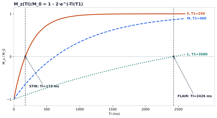
Schematic teaching values, not universal clinical constants: TI = T1·ln 2 assumes full equilibrium before a perfect 180° inversion — finite TR, imperfect inversion and sequence implementation shift the clinical null time; T1 depends on field strength and tissue state; actual image contrast also involves T2 weighting, readout and magnitude reconstruction. The figure explains the nulling mechanism; it does not predict clinical contrast. Pathological fluids need not share the schematic fluid’s T1.
Fat suppression is not one technique
STIR is one route to a fat-suppressed image. Spectral fat-suppression methods use the frequency difference between fat and water, while Dixon methods acquire and combine signals to estimate water and fat components. These approaches have different assumptions and sensitivities; none changes the central lesson that the resulting image is defined by its preparation, timing, sampling, and reconstruction. In particular, STIR’s null is tied to a T1 range, so changing field strength, tissue state, or sequence timing can change how closely the chosen TI suppresses the intended component.
The practical consequence is that a label such as “fat-suppressed” is not enough to identify the measurement conditions. Record the suppression method and its timing when comparing images or quantitative measurements. The next preparation changes the target from relaxation to motion: diffusion and ADC as model-based measurements.
Sources
- Abi Berger, “Magnetic resonance imaging” — pulse-sequence principles and relaxation-based contrast.
- Stanford Medicine Body MRI Research Group, MRI Physics Education Materials — public visual orientation on inversion recovery, fat suppression, and contrast mechanisms.
- Ridgway JP. Cardiovascular magnetic resonance physics for clinicians: part I. J Cardiovasc Magn Reson. 2010;12:71. doi:10.1186/1532-429X-12-71. (Full text)
- Fischer T, El Baz Y, Waelti S, et al. Short tau inversion recovery (STIR) after intravenous contrast agent administration obscures bone marrow edema-like signal on forefoot MRI. Skeletal Radiol. 2022;51:573-579. doi:10.1007/s00256-021-03852-2. (Full text)
- Lins CF, Salmon CEG, Nogueira-Barbosa MH. Applications of the Dixon technique in the evaluation of the musculoskeletal system. Radiol Bras. 2021;54:33-42. doi:10.1590/0100-3984.2019.0086. (Full text)
© 2026 Adrian Tkacz · BioMedical-IT
Original text and figures are licensed under the Creative Commons Attribution-NonCommercial 4.0 International License (CC BY-NC 4.0), unless otherwise noted. Please credit Adrian Tkacz / BioMedical-IT and link to the original source. Third-party materials retain their respective rights and licenses.